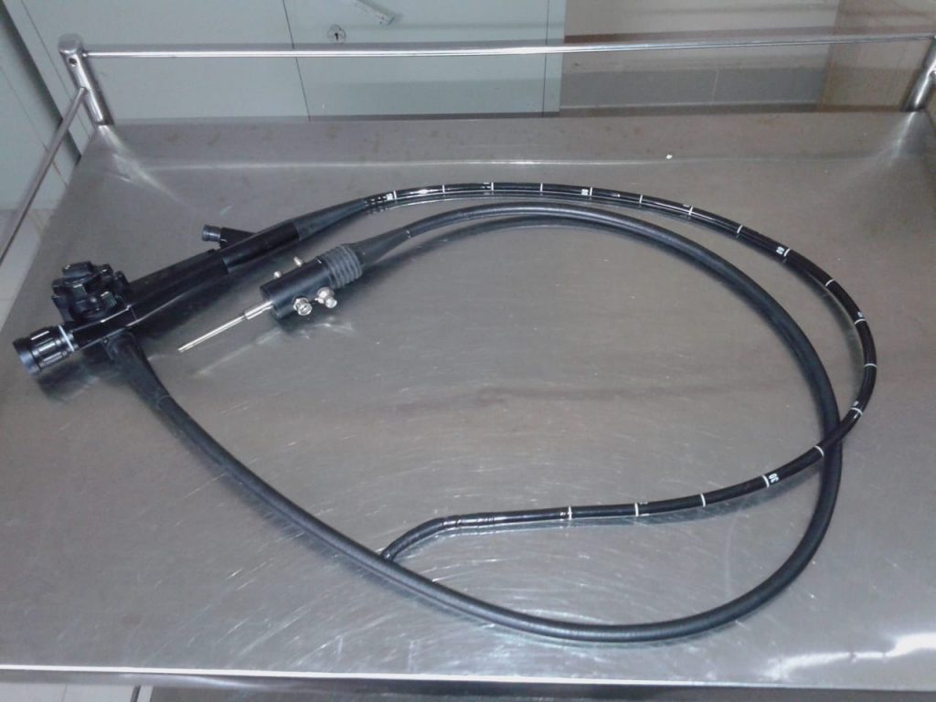
Endoscopy refers to various invasive procedures to examine the interior organs (such as kidney) or a cavity (such as mediastinum) of the body using an endoscope. Either for diagnostic and / or therapeutic purposes. The medical equipment used for examining the internal body cavities, called an endoscope, that may be roughly 1.5 meters or longer in some cases like a colonoscope. A cord connects the endoscope to the video processor, and contains the light guide, electrical connectors, and conduits for air, water, and suction. Details of some of the most common types of endoscopy procedures, the body parts examined and the scopes used can be found here – https://www.primedeq.com/blog/what-is-endoscopy-what-are-the-different-organs-examined-using-an-endoscope/ What are the key components of an endoscopy system? How does an endoscope work? Read on to know more.
What are Endoscopes and how are they used?
Endoscopes are diagnostic medical equipment, with a long tube, connected to a camera/ video camera. A light source is used to illuminate the area of interest, so that the camera can capture clear images. A fibre-optic cable system is connected from the light source and housed in the long insertion tube. Apart from the fibre-optic cables, the insertion tube also carries air/water channel, angulation guide wires and instrument channel.
The endoscope captures the required image and is transmitted to an image/ video processer that processes the images. The images are displayed on a HD medical grade monitor for real-time viewing by the physician, so as to perform appropriate diagnosis and procedure.
So what are the key components of an endoscopy system? How does an endoscope work?
Key components of an Endoscopy System
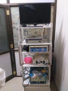
The basic endoscopy system includes: an endoscope, light source, image/video processing unit, medical grade monitor and suction system. The entire system is usually housed as a stack with a power supply source.
Endoscope
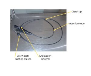
An endoscope is a medical equipment used to examine an internal organ like stomach or intestines. It has a small high definition camera at one end (called the distal end) of a long tube that can be inserted into the patient’s body through the nearest orifice, such as the mouth or anus. The rest of the control mechanism, image processing and all other supporting components are connected at the other end of the tube. The key components of the endoscope are:

- Insertion tube
- Distal tip
- The Objective Lens
- Angulation Control
- Air, water & suction valves
Insertion tube
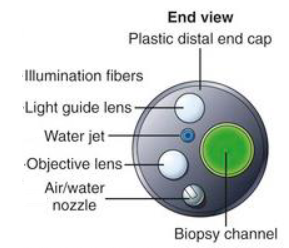
The insertion tube includes the following four main parts:
- Objective lens and image sensor,
- Light guides that bring light from the light source through the endoscope,
- Instrument channel outlet where instruments can be pushed in and taken out and suction,
- Nozzle for feeding water and air.
The objective lens is typically a super-wide-angle lens in order to visualize a large area of tissue at one time. In order to view tumor tissue in a more detailed manner, some endoscopes have an optical zoom feature. They also support high-definition video displays.
Light guide fibre bundles conduct light from the external light source through the endoscope to illuminate body cavities. Instruments may be pushed in and out of the instrument channel for harvesting tissue (biopsy), removing tumors, cauterizing bleeding lesions, etc. The nozzle on the distal tip is used to clean the lens with water and expand body cavities by insufflating them with air.

Distal End
The distal end of an endoscope contains the Optics that capture the endoscopic images, an instrument channel for delivery of instruments and removal of tissue and suction. The air/water channel directs water across the outer surface of the objective to clear debris or air to insufflate and expand the organ being examined.
Angulation control
The angulation control knobs for manipulating the distal end and the bending section contains a set of hinges that allow the distal tip to deflect at large angles as high as 270°, a lock for maintaining distal tip deflection, air/water and suction valves and remote switches for freezing, capturing, and storing images. The angulations are produced by a set of guide wires that deflect the distal end in four directions – up, down, left, and right. See more details on components of an endoscopy system – https://uomus.edu.iq/img/lectures21/MUCLecture_2023_1120947.pdf
Light Source
The cold light source, halogen, xenon or LED may be used for endoscopy. The physicians perform the investigation or biopsy while watching the image on a monitor screen instead of through-the-lens viewing.
Image processor
Image processor handles the process of recording, managing, and editing high-resolution endoscopic images (video and still images).
Monitor
An HD medical grade monitor (15” – 32”) is part of the system for high quality images to be viewed during the procedure.
Trolley

A trolley is typically used to house the entire ‘stack’ of endoscopy equipment or components of an endoscopy system. The trolley or mobile video cart generally has a drawer and three shelves to hold all the equipment and related instruments. The upper shelves have a tilt adjustment and used for supporting the video monitor. The trolley is equipped with an electrical supply points on the rear side. The light source, video system & recorder and the insufflator can all be placed in mobile trolley. The trolley or cart is equipped with locking brakes and wheels.
Popular endoscopy equipment brands are Olympus, Pentax, Fujifilm etc.
Also read
—————————————————————————————————————————
PrimedeQ is an e-Marketplace for buying, selling, renting, servicing and spares of medical equipment. We offer all types of used / refurbished medical equipment , including endoscopes, OT equipment, Laparoscopic Equipment, Lab equipment, X-Ray, TMT, ECG, anesthesia machine, ultrasound machines etc. We offer microdebrider, laparoscope , endoscope and ultrasound probe repair services etc.
https://in.linkedin.com/in/shanthi-mathur-ab07838
